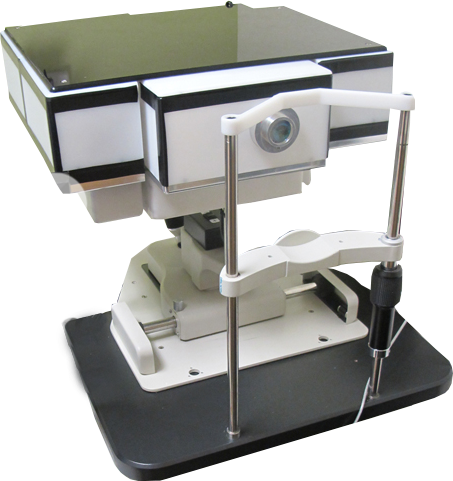
Aeon Imaging has developed the Laser Scanning Digital Camera (LSDC), a low-cost, easy to use device which records images of the back of the eye. The LSDC is a confocal line-scanning laser ophthalmoscope, protected by US patents and international patents pending.
The LSDC is not an FDA approved device. Please contact us if you'd like to use the LSDC for research.

Capabilities
Multiply Scattered Imaging Mode
Aeon's novel use of the CMOS electronic rolling shutter means that the LSDC can finely adjust the offset of its confocal aperture with respect to the illumination light on the retina electronically in real-time. With a confocal aperture offset, the LSDC records images composed of multiply scattered light from the deeper layers of the retina, while rejecting the much stronger direct backscatter from superficial layers. The result is increased sensitivity to scattering defects such as drusen and to the presence of edema.
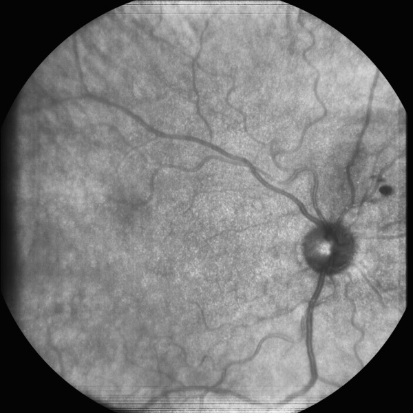
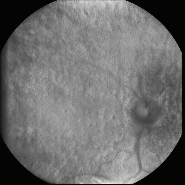
Retinal images taken of a 60 year old Caucasian female subject with dry age-related macular degeneration and widespread hard drusen are shown below. A 20-frame averaged standard LSDC image is shown on the left, and a 20-frame averaged multiply scattered mode on the right. Standard and multiply scattered image frames were alternately acquired.

Confocal LSDC image, showing typical superficial detail of the retina. The strong superficial back scattering masks the structure from the deeper layers in the retina.

Multiply scattered light image of the same eye. Dozens of small scattering disruptions (drusen) appear, which are a clinical hallmark of age-related macular degeneration (AMD).
Refraction Using Structured Illumination Mode
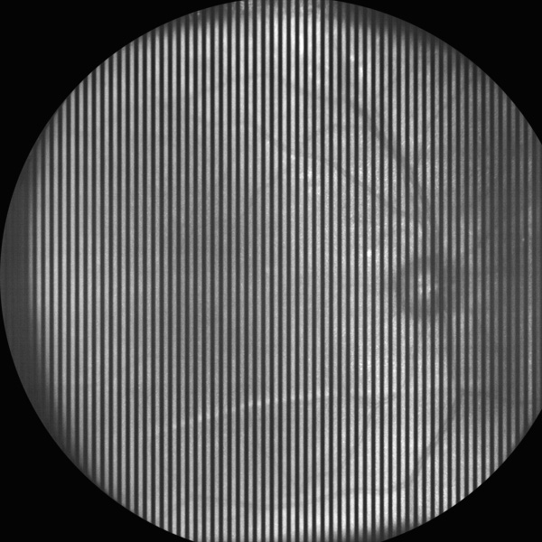
The LSDC's illumination source is more commonly used in telecommunication applications that require intensity modulation. In structured illumination mode, the source is modulated while imaging, producing a sequence of stripes across the image. The contrast or amplitude of the stripes measured through-focus provides a localized point-spread function. When measured near the fovea, this mode provides auto-focus; when combined with peripheral point-spread function measurements, the topography of the focused light on the retina can be reconstructed. Common uses for topography include modeling peripheral refraction and detecting retinal thickening commonly associated with eye disease.

LSDC image of a 23 year old Caucasian male with structured illumination. Tip: Try looking at the image up close and farther away. At farther distances, your eye will better average out the stripes, providing a clearer view of the retina."
Funding support for the development and evaluation of the LSDC has been provided by the National Institutes of Health Small Business Innovation Research program under grants R44EY020017 and R44EY018772. Matching grant support has also been provided by the Indiana Economic Development Corporation.
